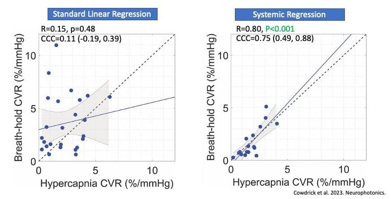
Cerebrovascular reactivity (CVR), i.e., the ability of cerebral blood vessels to dilate or constrict in response to changes in blood oxygen content or neuronal demand, is a biomarker of vascular health. CVR assessment usually involves administration of a controlled vasoactive stimulus so the reactive ability of the brain can be easily observed. The traditional way of doing this requires patients to undergo an MRI while performing what is called a “hypercapnia challenge” where they inhale carbon dioxide. In pediatric patients or after sever brain injury, this protocol is often infeasible or contraindicated. In recent work now out in Neurophotonics, Kyle R. Cowdrick et al. explored the application of Diffuse Correlation Spectroscopy (DCS) for CVR assessment in a cohort of healthy adults across multiple, more tolerable, experimental paradigms vs. a gold standard hypercapnia challenge. Specifically, they compared CVR calculated from DCS measurements taken from subjects breathing normally at rest or during a timed breath-hold challenge with hypercapnia. They found that applying general linear models to minimize influence of systemic hemodynamics on the brain signal measured with DCS improved the agreement between these more tolerable assessment methods and the gold-standard. This promising result suggests that DCS coupled with a milder vasoactive stimulus can allow CVR assessment in previously inaccessible patient groups.
