Monthly Archives: March 2023

New paper out on the influence of oversimplifying the head anatomy when using diffuse correlation spectroscopy to measure cerebral blood flow

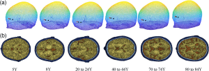
In our latest work in Neurophotonics, Hongting Zhao determines the influence of oversimplifying the head geometry on brain blood flow estimated with diffuse correlation spectroscopy (DCS). Due to the noninvasive nature of  DCS measurements, light must pass through extracerebral layers (i.e., skull, scalp, and cerebral spinal fluid) before detection at the tissue surface. To minimize the contribution of these extracerebral layers to the measured signal, an analytical model has been developed that treats the head as a series of three parallel and infinitely extending slabs (mimicking scalp, skull, and brain). The three-layer model has been shown to provide a significant improvement in cerebral blood flow estimation over the typically used model that treats the head as a bulk homogenous medium. However, the three-layer model is still a gross oversimplification of the head geometry that ignores head curvature, the presence of cerebrospinal fluid (CSF), and heterogeneity in layer thickness. Using Monte Carlo modeling in a four-layer slab medium and a three-layer sphere medium to isolate the influence of CSF and curvature, respectively, we found both head curvature and failing to account for CSF lead to significant errors in the estimation of cerebral blood flow. However, the effect of curvature and CSF on relative changes in blood flow is minimal. In sum, these findings suggest that the three-layer model holds promise for improving estimation of relative changes in cerebral blood flow; however, estimations of absolute cerebral blood flow with the approach should be viewed with caution given that it is difficult to account for appreciable sources of error, such as curvature and CSF.

Age-averaged MRI templates. (a) 3D mesh of the contour of each atlas along with the source (red) and detectors locations (black). (b) Axial view of the plane where source and detectors were placed (scalp in dark blue, skull in light blue, CSF in brown, and brain in yellow).

New paper out on blood flow biomarkers in subarachnoid hemorrhage

Check out our latest paper in Frontiers in Neurology. One of the common complications of non-traumatic subarachnoid hemorrhage (SAH) is secondary stroke known as delayed cerebral ischemia (DCI). Intrathecal (IT) administration of nicardipine, a calcium channel blocker, upon detection of large-artery cerebral vasospasm holds promise as a treatment that reduces the incidence of DCI. However, there are currently no reliable methods for determining of treatment is working in individual patients.

Image showing SAH patient with DCS sensor attached to the forehead

In this observational study, Sathialingam et al. prospectively employed a non-invasive optical modality called diffuse correlation spectroscopy (DCS) to quantify the acute microvascular cerebral blood flow response to intrathecally administered nicardipine in 20 patients with medium-high grade non-traumatic SAH, with the goal of assessing DCS-measured cerebral blood flow response as a potential biomarker of treatment efficacy. We observed that, on average, blood flow increased significantly with time post-treatment. However, individual patient responses fell into two distinct classes: (1) those showing no significant change in blood flow and (2) those showing a pronounced increase after treatment with IT nicardipine. The incidence of DCI was 5 out of 6 in Class 1 and 1 out of 13 in Class 2 (p < 0.001). These results suggest that the acute (<90 min) DCS-measured brain blood flow response to IT nicardipine is a biomarker of treatment efficacy, which could be used to identify non-responders in time to initiate alternative therapies to prevent secondary stroke after subarachnoid hemorrhage.